Revolutionizing healthcare and human-machine interfaces with wireless, high-resolution muscle tracking
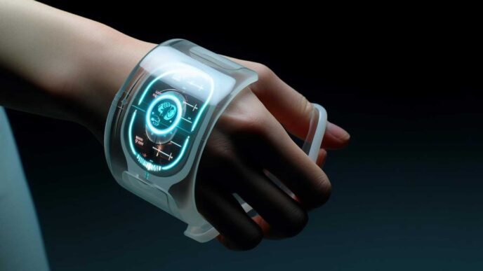
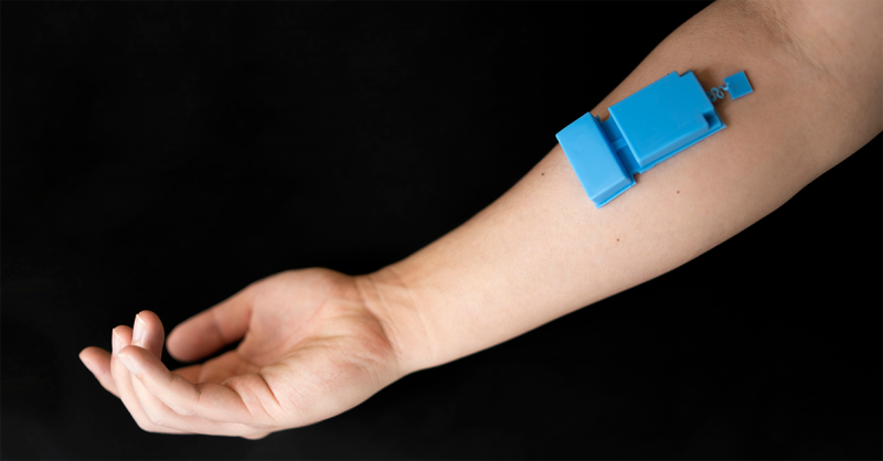
Engineers at the University of California San Diego have developed a groundbreaking wearable ultrasound device. This innovative technology enables long-term, wireless monitoring of muscle activity, opening new horizons in healthcare and human-machine interfaces. Designed to adhere to the skin with a layer of adhesive and powered by a battery, the device provides high-resolution tracking of muscle function without invasive procedures.
Key Features of the Wearable Ultrasound Device
• Wireless and Compact: The device operates without wires, enhancing comfort and mobility.
• High-Resolution Muscle Monitoring: Offers detailed insights into muscle function by penetrating deep tissues.
• Non-Invasive Application: Sticks to the skin using adhesive, eliminating the need for invasive procedures.
• Long-Term Use: Suitable for continuous monitoring during daily activities.
• AI Integration: Utilizes an artificial intelligence algorithm for precise muscle activity mapping.
The Team Behind the Innovation
The project was led by Professor Sheng Xu, a Jacobs Faculty Scholar in the Aiiso Yufeng Li Family Department of Chemical and Nano Engineering at UC San Diego. The research was a collaborative effort with Professor Jinghong Li, a pulmonologist and intensive care specialist at UC San Diego Health. Professor Joseph Wang, a distinguished professor in the same department, also co-authored the study.
Published Research
The team’s work was published on October 31 in Nature Electronics, highlighting the significance of this advancement in muscle monitoring technology.
Applications in Healthcare
Monitoring Respiratory Health
In tests, the device was worn over the rib cage to monitor diaphragm motion and thickness. This is crucial for assessing respiratory health.
• Benefits:
• Supports patients with respiratory conditions.
• Aids individuals reliant on mechanical ventilation.
• Provides continuous, real-time data on diaphragm activity.
Quote:
“By tracking diaphragm activity, the technology could potentially support patients with respiratory conditions and those reliant on mechanical ventilation,” said Professor Joseph Wang.
Advancements Over Traditional EMG
The wearable ultrasound offers a promising alternative to electromyography (EMG), which involves applying metal electrodes to the skin. EMG has limitations such as:
• Low Resolution: Difficulty isolating specific muscle fibers.
• Weak Signals: Signals from multiple muscle fibers blend together.
• Invasiveness: Less comfortable for long-term use.
The new device overcomes these challenges by providing:
• High-Resolution Imaging: Penetrates deep tissues for detailed muscle insights.
• Compact Design: Comfortable for continuous wear.
• Wireless Functionality: Enhances patient mobility.
Human-Machine Interface Potential
Researchers successfully used the device on the forearm to capture hand and wrist muscle activity. This enabled:
• Control of a Robotic Arm: Demonstrating applications in prosthetics and robotics.
• Playing a Virtual Game: Showcasing potential in interactive gaming and rehabilitation.
Technical Innovations
Device Composition
The device is housed in a flexible silicone elastomer casing and consists of three main components:
1. Single Ultrasound Transducer: Sends and receives ultrasound waves.
2. Custom-Designed Wireless Circuit: Controls the transducer, records data, and transmits it wirelessly.
3. Lithium-Polymer Battery: Powers the system for at least three hours.
Key Innovations
• Single Transducer Efficiency: Uses one ultrasound transducer to effectively sense deep tissues.
• Intensity-Controlled Ultrasound Waves: Enhances signal quality and depth penetration.
• High Spatial Resolution: Achieves precise isolation of specific muscle movements.
AI Algorithm Integration
The researchers developed an artificial intelligence algorithm that:
• Maps Radiofrequency Signals: Translates signals into corresponding muscle distributions.
• Identifies Specific Hand Gestures: Achieves high accuracy and reliability in gesture recognition.
• Enhances Human-Machine Interaction: Improves control mechanisms for prosthetics and interfaces.
Advantages Over Existing Technologies
• Continuous Monitoring: Suitable for long-term wear during daily routines.
• Non-Invasive and Comfortable: Flexible and adherent design enhances user comfort.
• Detailed Muscle Analysis: Provides deeper insights compared to EMG.
• Wireless Data Transmission: Eliminates the need for cumbersome wires.
Future Implications
The wearable ultrasound device holds significant promise for:
• Medical Diagnostics: Early detection and monitoring of muscular and respiratory conditions.
• Rehabilitation: Assisting in physical therapy by providing real-time muscle activity feedback.
• Prosthetics: Enhancing control over prosthetic limbs through precise muscle signal detection.
• Human-Machine Interfaces: Advancing interactive technologies in gaming and virtual reality.
Conclusion
The development of this wearable ultrasound device marks a significant leap forward in muscle monitoring technology. By combining high-resolution imaging, wireless functionality, and AI algorithms, the engineers at UC San Diego have opened new pathways in healthcare and human-machine interfaces.
This innovative approach addresses the limitations of traditional methods like EMG, providing a more detailed, comfortable, and practical solution for continuous muscle monitoring.
Learn More
For more detailed information on this groundbreaking research, visit the UC San Diego News Center.
Meta Description: